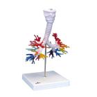
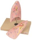
Model Ct Bronchial Tree, with Larynx and Transparent Lung, Dimensions: 22x18x37 cm, Weight: 1.905 kg, created on the basis of computer tomography data of a human, The larynx with hyoid bone and epiglottis and the trachea with primary and lobar
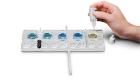
Human Anatomy Model, The knee bone is connected to the arm bone, Eight awesome activities explore the human body from the inside out, including optical camera, tooth check-in, taste test, handy thermometer, pumping heart, lung simulation, torso model and DNA design
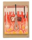
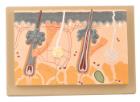
Comprehensive model 300 times, enlarged vertical section showing the three layers, sebaceous and sweet glands, the hair follicles, erector muscles, arteries, nerves and veins. With key card
Enlarged 70 Times, full size, shows section through three layers of the hair covered skin of the head. Also shows hair follicles with sebaceous, sweat glands, receptors, nerves and vessels.
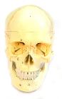
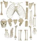
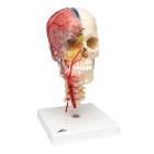
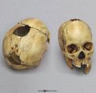
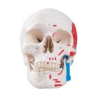
Each part on the skull is numbered. Jaw is spring loaded. Skull cap is removable for further interior inspection. Included with the model is a sheet with each numbers name for identification.
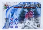
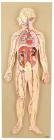
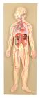
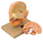
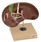
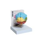
This half-size relief model helps in understand the circulation of blood in body through brain, heart, lung, liver, spleen, kidneys and partial skeleton. Mounted on base. Numbered with key card.
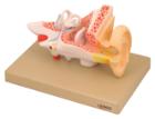
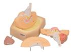
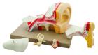
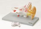
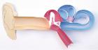
Enlarged approximately 4 times. The petros portion of the temporal bone section of the auditory canal are removable, with labyrinth can be taken out and opened. The tympanic membrane with malleus and incus can be removed in 5 parts.
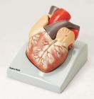
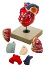
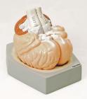
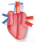
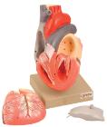
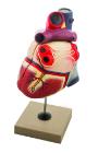
Enlarged. Sectioned so that both ventricles and atria open to expose the valves. Large blood vessels near the heart and musculature of the heart are shown. Separates into 7 parts.
Skull Trauma Set of Six Fragments model, with Shotgun Wounds, aping defect on the occiput involves the lambda; internal beveling is palpable along the edges of the wound, sagittal suture, Size: 20.1L x 13W x 17.3H (cm)
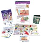
Human Body III: Maintaining Life Curriculum Learning Module provides Complete set of digital and hands-on resources, includes: Flip Chart Set; Student Learning Guide; Vocabulary Card Set; Curriculum Mastery Game; 1 Interactive Multimedia Lesson; and Hands-On Pathogens Lab.
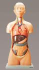
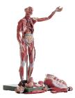
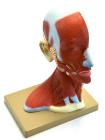
Life size model showing the outer superficial muscles, vessels, nerves and head with muscles on one side. On the outer side details of median section such as brain, mouth, larynx are shown. Includes key card.
Sectioned through the ventricles and auricles. The bicuspid and tricuspid semilunar and sigmoid valves are shown. Separates into 3 parts. Mounted on base
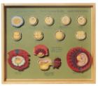
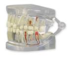
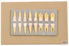
Enlarged approximately 2 times, set of 16 teeth cast in break resistant material having accurate anatomical details. Complete set as in half of upper & lower jaw
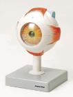
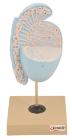
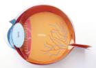
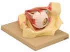
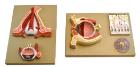
Model shows both sides of an eye, enlarged 5 x. One side of the model shows the eye socket with a sagittal cutaway and the background to the eye and the electron microscopic fine structure of the retina are shown separately.
Life size model dissectible in 2 parts. The anterior heart wall can be removed to show the left and right ventricles and atria as well as the tricuspid, pulmonary, mitral and aortic valves. Mounted on base.